
The laboratory of Salahedin Ayuobi Hospital is equipped with modern equipment and has experienced specialists who provide the following services to the respectable patients every day.
CBC,FBS,Urea,Cr,Na,Bili-T,SGPT (ALT),FBS,Amylase,TG,Uric Acid,PTT,ABO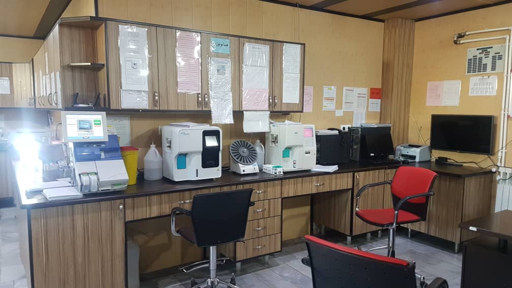
The CT scan machine is spiral multi-slice 2 detector
Heavy weight patients (up to 125 kg) can use this device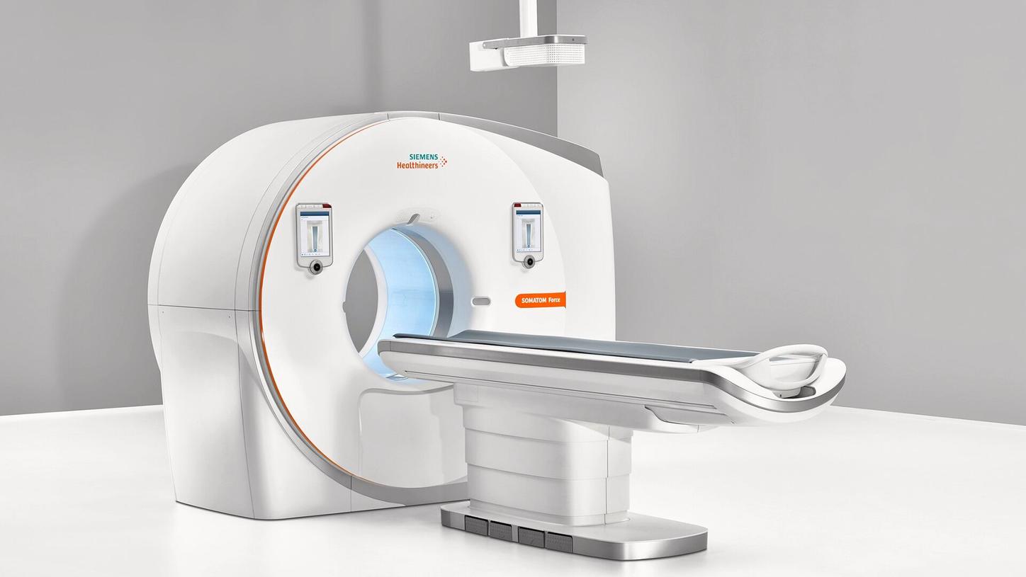


An electrocardiogram is a painless, noninvasive way to help diagnose many common heart problems. A health care provider might use an electrocardiogram to determine or detect: Irregular heart rhythms (arrhythmias) If blocked or narrowed arteries in the heart (coronary artery disease) are causing chest pain or a heart attack Whether you have had a previous heart attack. How well certain heart disease treatments, such as a pacemaker, are working.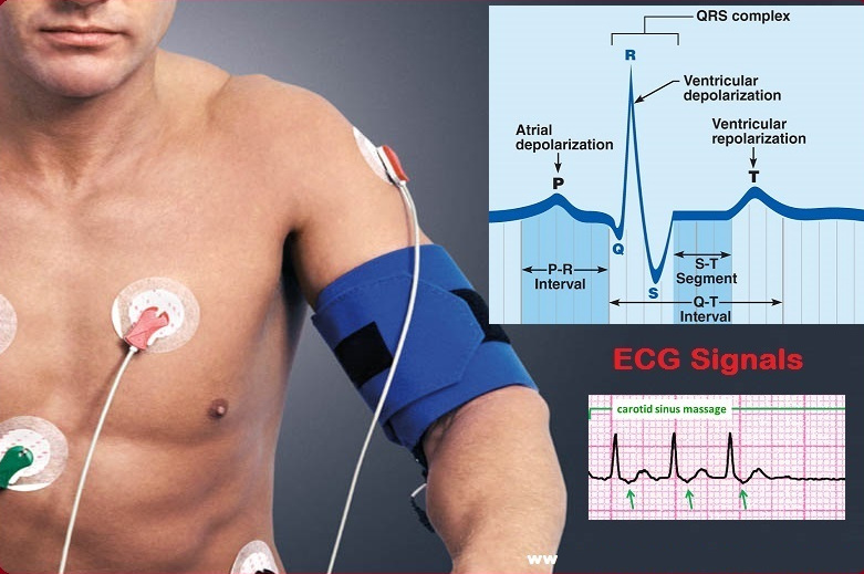
ectromyography (EMG) measures muscle response or electrical activity in response to a nerve’s stimulation of the muscle. The test is used to help detect neuromuscular abnormalities. During the test, one or more small needles (also called electrodes) are inserted through the skin into the muscle. The electrical activity picked up by the electrodes is then displayed on an oscilloscope (a monitor that displays electrical activity in the form of waves). An audio-amplifier is used so the activity can be heard. EMG measures the electrical activity of muscle during rest, slight contraction and forceful contraction. Muscle tissue does not normally produce electrical signals during rest. When an electrode is inserted, a brief period of activity can be seen on the oscilloscope, but after that, no signal should be present.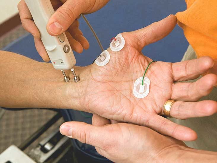
Spirometry (spy-ROM-uh-tree) is a common office test used to assess how well your lungs work by measuring how much air you inhale, how much you exhale and how quickly you exhale.
Spirometry is used to diagnose asthma, chronic obstructive pulmonary disease (COPD) and other conditions that affect breathing. Spirometry may also be used periodically to monitor your lung condition and check whether a treatment for a chronic lung condition is helping you breathe better.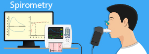
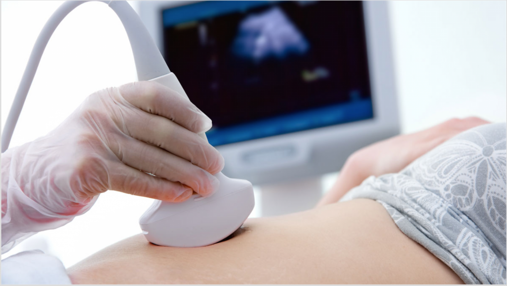
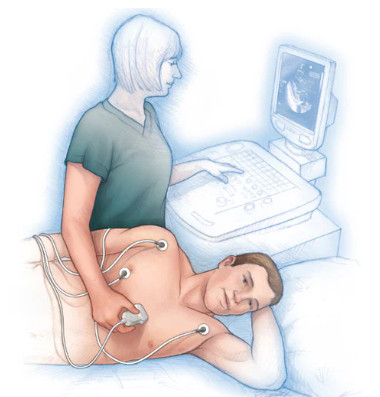
An echocardiogram uses sound waves to produce images of your heart. This common test allows your doctor to see your heart beating and pumping blood. Your doctor can use the images from an echocardiogram to identify heart disease. Depending on what information your doctor needs, you may have one of several types of echocardiograms. Each type of echocardiogram involves few, if any, risks.
 |
 |
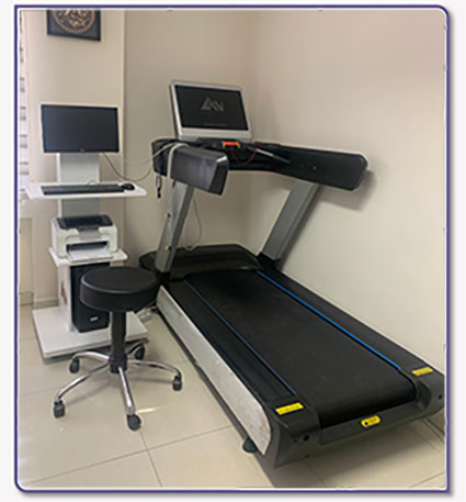
A stress test, also called an exercise stress test, shows how your heart works during physical activity. Because exercise makes your heart pump harder and faster, an exercise stress test can reveal problems with blood flow within your heart. A stress test usually involves walking on a treadmill or riding a stationary bike while your heart rhythm, blood pressure and breathing are monitored. Or you'll receive a drug that mimics the effects of exercise.